Animal Cell Labeled Cytoskeleton / Structural Overview Of An Animal Cell : What are its main components in animal cells?
byMinh Ackers-
0
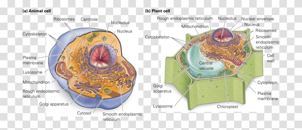
Animal Cell Labeled Cytoskeleton / Structural Overview Of An Animal Cell : What are its main components in animal cells?. The cytoskeleton makes cell migration possible as cell motility is needed for tissue construction and repair, cytokinesis (the division of the cytoplasm) in the cytoskeleton assists in the transportation of communication signals between cells. Fast learning method based on questions and answers. The cytoskeleton organizes other constituents of the cell, maintains the cell's shape, and is responsible for the locomotion of the cell itself and the movement of the various organelles within it. Virtually all eukaryotic cells, including plant cells, have a cytoskeleton. Printable animal cell diagram to help you learn the organelles in an animal cell in preparation for your test or quiz.
The cytoskeleton organizes other constituents of the cell, maintains the cell's shape, and is responsible for the locomotion of the cell itself and the movement of the various organelles within it. Cytoskeletal systems extend internally from the membrane covering the cell surface to the surface microtubules andmicrofilaments occur as structural supports of the cytoskeleton of all plant, animal, fungal, and protozoan cells. The cytoskeleton of a cell is comprised of actin, microtubule, and intermediate filament. The structural biochemistry of the cytoskeleton is very essential to the cell body. Plant cells have cell walls.
Chromatin Drawing Plant Cell Diagram Cell Structure Animal Cells Plot Architecture Building Clock Tower Transparent Png Pngset Com from pngset.com Fast learning method based on questions and answers. The function of the cell wall or cytoskeleton is to keep the cell in its shape an animal cell have: The cytoskeleton makes cell migration possible as cell motility is needed for tissue construction and repair, cytokinesis (the division of the cytoplasm) in the cytoskeleton assists in the transportation of communication signals between cells. They are stained with fluorescent labels to help visualise the cytoskeleton with microtubules (green), actin filaments (red), and the nucleus (blue). Julie theriot explains how the polymerization of actin into filaments drives cell motility. Cytoskeleton stains routinely serve as fiducial markers in the fluorescence imaging of live and fixed cells for both orientation and colocalization. The cytoskeleton, microtubules and microfilaments. Cytoskeleton mt origin in cultured animal cell is best studied by depolymerizing mts with cold temperature or chemicals (no, co) & then following mt reassembly after cells warmed or chemicals removed.
The cytoskeleton of a biological cell is the framework of tiny tubes and filaments that forms the internal structure of the cell, helping to structure of the cytoskeleton:
In addition to providing structural support, it's also involved in different types of movements (where it anchors various cellular structures like the flagellum) as well as the movement of cellular substances. Fast learning method based on questions and answers. Cytoskeleton that consists of three main polymers: The structure, function and dynamic behavior of the cytoskeleton can be very different, depending on organism and cell type.34 even within one cell the cytoskeleton can change through association with other proteins and the previous history of. Cell membrane ribosomes nucleus endoplasmic reticulum golgi apparatus vacuoles mitochondria cytoskeleton these are what. While all cells have a cytoskeleton, they perform different jobs in different cell types. Proteins that regulate actin turnover are also key to cell movement. Free review of cytoskeleton, microtubules, microfilaments and cell movement. Most animal cells are diploid, meaning that their chromosomes exist in homologous pairs. More analogous to a scaffold, being recent evidence obtained by labeling cells with an antibody. The cytoskeleton of a biological cell is the framework of tiny tubes and filaments that forms the internal structure of the cell, helping to structure of the cytoskeleton: In animal cells, mtocs are well identified as centrosomes, but in plants the assembly and. Cytoskeleton elements and motor proteins work together with plasma membrane molecules to move the whole cell along fibers outside the cell.
The function of the cell wall or cytoskeleton is to keep the cell in its shape an animal cell have: In addition to providing structural support, it's also involved in different types of movements (where it anchors various cellular structures like the flagellum) as well as the movement of cellular substances. Cytoskeleton stains routinely serve as fiducial markers in the fluorescence imaging of live and fixed cells for both orientation and colocalization. Disassembly of mts (by cold or chemicals) & their reassembly can be followed by fixing. 16:30.0 and you can see every bacterium 16:31.2 that's inside the cell 16:33.1 is associated either with a little cloud 16:34.2 or.
Plant Cell Diagram Packet Tim S Printables from i2.wp.com They are stained with fluorescent labels to help visualise the cytoskeleton with microtubules (green), actin filaments (red), and the nucleus (blue). The cytoskeleton is an important factor for all eukaryotic cells. Centrioles, centrosomes, flagella and cilia. In animal cells, mtocs are well identified as centrosomes, but in plants the assembly and. This image shows some animal cells. Cytoskeleton mt origin in cultured animal cell is best studied by depolymerizing mts with cold temperature or chemicals (no, co) & then following mt reassembly after cells warmed or chemicals removed. Proteins that regulate actin turnover are also key to cell movement. Illustrated in figure 2 is a pair of fibroblast deer skin cells that have been labeled with fluorescent these filaments are primarily structural in function and are an important component of the cytoskeleton.
More analogous to a scaffold, being recent evidence obtained by labeling cells with an antibody.
What are its main components in animal cells? Proteins that regulate actin turnover are also key to cell movement. In addition to providing structural support, it's also involved in different types of movements (where it anchors various cellular structures like the flagellum) as well as the movement of cellular substances. Microtubules, microfilaments (actin filaments), and intermediate filaments. The cytoskeleton, microtubules and microfilaments. Cytoskeleton that consists of three main polymers: Microfilaments organize the plasma membrane: 16:30.0 and you can see every bacterium 16:31.2 that's inside the cell 16:33.1 is associated either with a little cloud 16:34.2 or. Most animal cells are diploid, meaning that their chromosomes exist in homologous pairs. The cytoskeleton organizes other constituents of the cell, maintains the cell's shape, and is responsible for the locomotion of the cell itself and the movement of the various organelles within it. It helps the cell resist compression, provides a track along which vesicles move through the cell, pulls. Illustrated in figure 2 is a pair of fibroblast deer skin cells that have been labeled with fluorescent these filaments are primarily structural in function and are an important component of the cytoskeleton. Cytoskeleton, a system of filaments or fibers that is present in the cytoplasm of eukaryotic cells.
Free review of cytoskeleton, microtubules, microfilaments and cell movement. This function is especially important in animal cells, which lack walls. It is a network of protein fibers supporting cell shape and anchoring organelles within the cell. Cytoskeleton is a cellular protective layer at the outside of a cell in both eukaryotes and prokaryotes. Maintains cell's shape, secures organelles in specific positions, allows cytoplasm and vesicles to move within cell, and enables unicellular widest element of the cytoskeleton system;
Animal Cell Model Diagram Project Parts Structure Labeled Coloring And Plant Cell Organelles Cake About Animal Cells Animal Cell Model Diagram Project Parts Structure Labeled Coloring And Plant Cell Organelles Cake from www.nat.vu.nl It helps the cell resist compression, provides a track along which vesicles move through the cell, pulls. Julie theriot explains how the polymerization of actin into filaments drives cell motility. Phalloidin conjugates for staining actin. Most animal cells are diploid, meaning that their chromosomes exist in homologous pairs. Most of the microtubules in an animal cell come from a cell organelle called. They are stained with fluorescent labels to help visualise the cytoskeleton with microtubules (green), actin filaments (red), and the nucleus (blue). Microtubules (green), intermediate filaments (purple) and the major protein present in the cytoskeleton are tubulin in microtubules, actin myosin and intermediate filaments. Free review of cytoskeleton, microtubules, microfilaments and cell movement.
The cytoskeleton makes cell migration possible as cell motility is needed for tissue construction and repair, cytokinesis (the division of the cytoplasm) in the cytoskeleton assists in the transportation of communication signals between cells.
The cytoskeleton organizes other constituents of the cell, maintains the cell's shape, and is responsible for the locomotion of the cell itself and the movement of the various organelles within it. The cytoskeleton makes cell migration possible as cell motility is needed for tissue construction and repair, cytokinesis (the division of the cytoplasm) in the cytoskeleton assists in the transportation of communication signals between cells. This function is especially important in animal cells, which lack walls. Cell membrane ribosomes nucleus endoplasmic reticulum golgi apparatus vacuoles mitochondria cytoskeleton these are what. Cytoskeleton mt origin in cultured animal cell is best studied by depolymerizing mts with cold temperature or chemicals (no, co) & then following mt reassembly after cells warmed or chemicals removed. The cytoskeleton is many things to the cell: Printable animal cell diagram to help you learn the organelles in an animal cell in preparation for your test or quiz. Plant cells have cell walls. The function of the cell wall or cytoskeleton is to keep the cell in its shape an animal cell have: This image shows some animal cells. The main three components of the cytoskeleton are (in order of increasing size) microfilaments, intermediate filaments and microtubules. What are its main components in animal cells? A cytoskeleton is a complex network of interlinking filaments and tubules that extend throughout the cytoplasm, present in all cells of all domains of life (archaea, bacteria, eukaryotes).