Animal Cell Cycle Labeled - Membrane And Organelle Dynamics During Cell Division Nature Reviews Molecular Cell Biology / Not all cells adhere to the classic cell cycle pattern in which a newly formed daughter cell immediately enters the preparatory phases of interphase, closely followed by the mitotic phase.
byMinh Ackers-
0
Animal Cell Cycle Labeled - Membrane And Organelle Dynamics During Cell Division Nature Reviews Molecular Cell Biology / Not all cells adhere to the classic cell cycle pattern in which a newly formed daughter cell immediately enters the preparatory phases of interphase, closely followed by the mitotic phase.. Destroy worn out damaged organelles. Animal cell diagram labeling labelled diagram. Animal gene functions required for cell cycle progression can be identified. Label both a plant and animal cell on a poster layout. Mitochondria, smooth and rough er, lysosomes, golgi apparatus, vacuoles, centrioles, ribosomes, cytoplasm, cell.
These events are tightly regulated and precisely timed, and can be grouped into two phases: In this video i'm going to draw labelled diagram of animal cell.in this video you will see the diagram of animal cell and it's labelling.this diagram of. Interphase and the mitotic (m) phase. In animal cells during prophase, microscopic bodies called centrioles begin to migrate to opposite sides of the cell. Cells grow at different rates in each of the different phases of the growth cycle and the calculated.
Cell Cycle Label from www.biologycorner.com In cells without a nucleus (prokaryotic), the cell cycle occurs via a process termed binary fission. Not all cells adhere to the classic cell cycle pattern in which a newly formed daughter cell immediately enters the preparatory phases of interphase, closely followed by the mitotic phase. Stem cell research dolan dna learning center. The cell cycle involves many repetitions of cellular growth and reproduction. In this phase, the cell increases in mass and organelle number in preparation for cell division. By george von dassow, university of oregon. In animal cells during prophase, microscopic bodies called centrioles begin to migrate to opposite sides of the cell. Include descriptions of what each part does.
Through corresponding conditional mutant animal cell lines defective in.
Visualize a cell's parts with this sturdy soft foam model. Chromosomes are stained with dapi (blue) and actin filaments are labeled with fluorescent phalloidin (yellow). Label both a plant and animal cell on a poster layout. In animal cells, cytokinesis results when a fiber ring composed of a protein called actin around the i. Animal cell diagram labeling labelled diagram. Mitochondria, smooth and rough er, lysosomes, golgi apparatus, vacuoles, centrioles, ribosomes, cytoplasm, cell. Where, prokaryotes are just bacteria and archaea to check if you have understood the cell parts, draw a blank animal cell diagram and try to fill in the different parts without referring to the labeled one given. Structure and support for the cell. There is a printable worksheet available for download here so you can take the quiz with pen and paper. Not all cells adhere to the classic cell cycle pattern in which a newly formed daughter cell immediately enters the preparatory phases of interphase, closely followed by the mitotic phase. One half has labeled parts: This is an online quiz called this animal cell needs labelling! Find diagrams of a plant and an animal cell in the science tab.
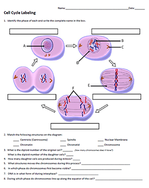
Using arrows and textables, label each part of the cell and describe its function. Cells on the path to cell in cells such as animal cells that lack cell walls, cytokinesis begins following the onset of anaphase. Interphase, prophase, metaphase, anaphase, and telophase. In animal cells during prophase, microscopic bodies called centrioles begin to migrate to opposite sides of the cell. Find diagrams of a plant and an animal cell in the science tab.
The Cell Cycle Biology For Majors I from s3-us-west-2.amazonaws.com The cell cycle is an ordered series of events involving cell growth and cell division that produces two new daughter cells. Are special vesicles in animal cells that contain enzymes. As cell cycle duration varies among cells in mammalian tissue culture cells, we asked whether their division asymmetry contributes to this variability. Cytokinesis occurs after mitosis and is different in plant and animal cells. Students label the image of a cell undergoing mitosis and answer questions about the cell cycle. Find diagrams of a plant and an animal cell in the science tab. These events are tightly regulated and precisely timed, and can be grouped into two phases: Through corresponding conditional mutant animal cell lines defective in.
Cell cycle tutorial from cells alive! cell death kuby immunology.
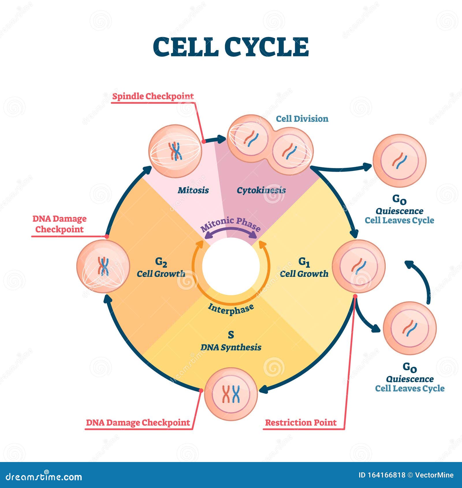
The structure of an animal cell, with labeled parts. With few exceptions (for example, red blood cells), all the cells of living things. Through corresponding conditional mutant animal cell lines defective in. The cell cycle is an ordered series of events involving cell growth and cell division that produces two new daughter cells. The cell cycle is the complex sequence of events by which cells grow and divide. Unlike the eukaryotic cells of plants and fungi, animal cells do not have a cell wall. Mitochondria, smooth and rough er, lysosomes, golgi apparatus, vacuoles, centrioles, ribosomes, cytoplasm, cell. Eukaryotic cells are larger, more complex, and have evolved more recently than prokaryotes. The cell cycle describes a sequence of reactions that results in the growth of the cell and replication of the genetic material to make two identical daughter cells. Visualize a cell's parts with this sturdy soft foam model. A more comprehensive reference on animal cell culture can be found in culture of animal cells: During development from stem to fully differentiated, cells in the body alternately divide (mitosis) and appear to be resting. Label both a plant and animal cell on a poster layout.
Stem cell research dolan dna learning center. By george von dassow, university of oregon. Chromosomes are stained with dapi (blue) and actin filaments are labeled with fluorescent phalloidin (yellow). These events are tightly regulated and precisely timed, and can be grouped into two phases: Using arrows and textables, label each part of the cell and describe its function.
Cell Cycle Stock Illustrations 1 815 Cell Cycle Stock Illustrations Vectors Clipart Dreamstime from thumbs.dreamstime.com By george von dassow, university of oregon. These events are tightly regulated and precisely timed, and can be grouped into two phases: It's the cell's brain, employing chromosomes to instruct other parts of the cell. Find diagrams of a plant and an animal cell in the science tab. Most of the cells size range between 1 and 100 micrometers and are visible only with the microscope. One half has labeled parts: One vital part of an animal cell is the nucleus. Cytokinesis occurs after mitosis and is different in plant and animal cells.
One half has labeled parts:
The main phases are shown: The cell cycle is an ordered series of events involving cell growth and cell division that produces two new daughter cells. By george von dassow, university of oregon. In eukaryotic cells, this process includes a series of four distinct phases. Animal cells are packed with amazingly specialized structures. Animal cell diagram labeling labelled diagram. Interphase is a very active phase of the cell cycle with many processes occurring in the nucleus and cytoplasm. Find diagrams of a plant and an animal cell in the science tab. Are special vesicles in animal cells that contain enzymes. One vital part of an animal cell is the nucleus. Aspects of dna replication, mitosis, and cytokinesis, instead concentrating. Visualize a cell's parts with this sturdy soft foam model. Science biology nutrient cycle carbon cycle.